Transdermal Transport
 Project Background: Human skin provides a two-way barrier that prevents potentially harmful chemicals or diseases from entering the body while slowing water as it exits the body. These barrier effects are mainly due to the outer-most layer of skin, called the stratum corneum (SC). The SC is composed of many corneocyte (dead cell) “bricks” in a lipid bilayer continuum “mortar”; in order to reach the bloodstream, any molecule on the surface of the skin must pass through the SC.
- Mechanical Extension (Stretching): Studies on the mechanical behavior of skin have shown that lipids, the permeable domain in skin, should stretch significantly more than the (relatively impermeable) corneocytes when skin is stretched. We are investigating the potential increase in skin permeability.
- Hydration: Although levels of skin hydration can alter the transdermal transport of toxins and drugs by a factor of 6 or more, possibly by altering transport pathways, current methods for in vitro testing do not account for effects of variable skin hydration. We are investigating the effects of hydration on overall transport rate and transport pathways for both hydrophilic and hydrophobic (lipophilic) molecules via experiments and finite element simulations.
Our Work:
- In Vitro Franz Cell Experiments: To test the permeability of skin in vitro at different hydration levels, we run permeation experiments on excised skin using Franz cells. Skin is placed between two chambers, one with a saturated drug solution and the other with no drug (water or buffer). Samples are collected at various time intervals and analyzed using HPLC (high-performance liquid chromatograph) or capillary electrophoresis (CE).
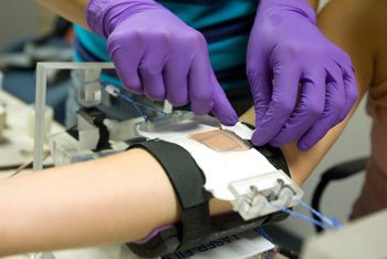
- In Vivo Tape-Stripping Experiments: To examine the effects of stretching on transport through skin, we conduct in vivo studies on human participants. Using a stretching apparatus designed by the research team, we apply either constant strain (fixed displacement) uniaxial extension or constant stress (fixed force) to the forearms of volunteers for the in vivo experiments. We place two drug-saturated patches on each volunteer’s forearm, one on the control (un-stretched) site and one on stretched skin. We then use a technique called tape stripping to remove several layers of the SC and analyze the results to compare transport across stretched vs. unstretched skin.
- Finite Element Modeling: We are using the finite element modeling (FEM) program COMSOL to run simulations of transport across the skin as the geometry (lipid and corneocyte dimensions) change with hydration or mechanical extension.
Click here to read an article about this work. To read more about the students involved in this project, click here. Interested in joining this research team? Please click here for more information.