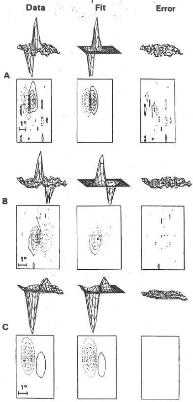
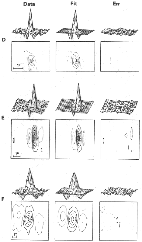
The spatiotemporal Gabor filter model for the V1 cells is supported by biological evidences from single cell recordings (Jones and Palmer 1987, Mclean and Plmer 1989). The two figures below show the comparisons of the response profiles of six V1 simple cells with the 2D Gabor models that best fit them. In all six cases, the left plot is the actual recording data, the middle one is the Gabor function that best fits the data, and the right one is the error, the difference between the real data and the Gabor model, which is quite small in all six cases.
The 3D spatiotemporal Gabor model is also supported by the single cells recordings.
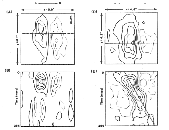
The figure below shows the spatiotemporal response profiles of two simple V1 cells.
First, the 2D spatial response profiles of the cells are shown in (A) and (D),
where the horizontal axis is ![]() and vertical one is
and vertical one is ![]() , with the time fixed at
, with the time fixed at
![]() miliseconds. Then the 2D spatiotemporal response profiles of the cells are
shown in (B) and (E), where the horizontal axis is still
miliseconds. Then the 2D spatiotemporal response profiles of the cells are
shown in (B) and (E), where the horizontal axis is still ![]() , the vertical one is
time
, the vertical one is
time ![]() , with the other spatial variable
, with the other spatial variable ![]() fixed at some constant position in
the receptive field indicated by the horizontal lines in (A) and (D). As can be
seen, the cell on the left (B) is not sensitive to motion, while the cell on the
right (E) is sensitive to rightward motion.
fixed at some constant position in
the receptive field indicated by the horizontal lines in (A) and (D). As can be
seen, the cell on the left (B) is not sensitive to motion, while the cell on the
right (E) is sensitive to rightward motion.