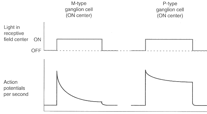
- They have large dentritic fields and large receptive fields. An M-cell is connected to a group of diffuse bipolar cells each of which is in turn connected with a group of receptors.
- They conduct action potentials more rapidly in the axon (optic nerve);
- The time course of their response (firing rate) only has a strong transient component without much sustained component;
- They are sensitive to the directions of visual motion.
- They are sensitive to low contrasts and they saturate when the the contrast is high;
- They are not sensitive to colors of the lights. They only have black and white center-surround receptive fields. [(ML)+ cetner/(ML)- surround, or (ML)- cetner/ (ML)+ surround.]
P-type ganglion cells constitute about 70 % the ganglion cell population. P-type cells have the following properties:
- They have smaller dendritic fields and smaller receptive field
than the M-type cells. In the fovea, a P-cell is connected to a single
midget bipolar cell, which in turn is directly connected to a single
cone (L or M, but not S).
Due to this architechture, the P-cells sample the visual field up to the full resolution of the core receptors (about 1 minute) for fine spatial resolution.
- They conduct action potentials at a slower speed in the axon (optic nerve);
- The time course of their response has sustained as well as transient components;
- They are more sensitive to the form and fine details of the visual stimuli;
- They respond poorly to low contrast but do not saturate at hight contrasts;
- They are sensitive to differences in the wavelength of light. Most of these P cells have color-opponent center-surround receptive fields for red-green (M+ center/L- surround, M- center/L+ surround, L+ center/M- surround, L- center/M+ surround). This is the first site where red and green are compared.


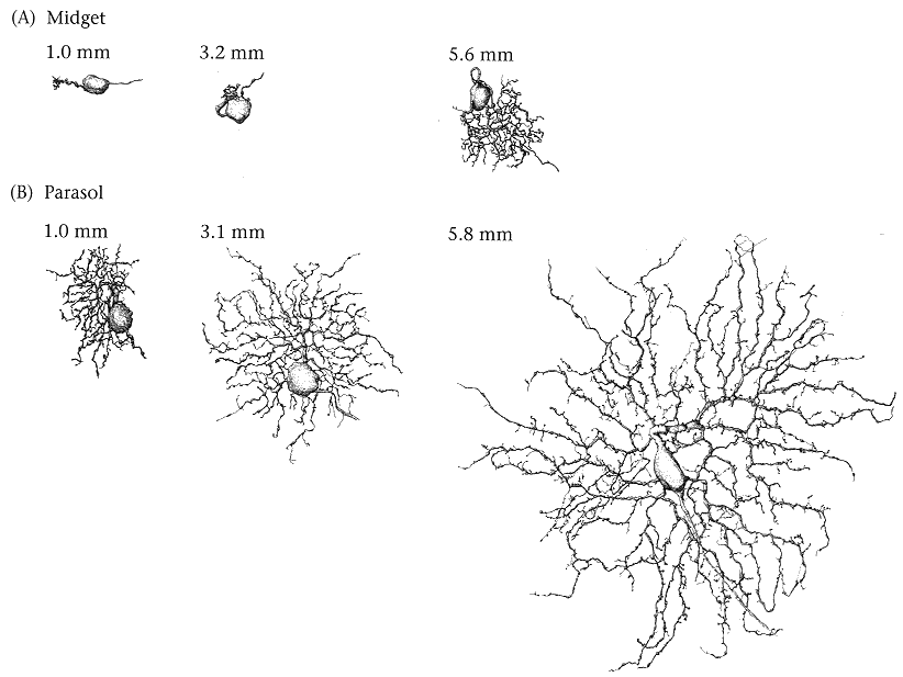
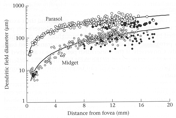
As one can see in this figure, the size of the dentrite tree for both midget and parasol cells increases as a function of the eccentricity, their distance to the fovea center. But parasol cells are always larger than midget cells at any eccentricity.


Their dendritic trees are stratified into two tiers near the inner and outer borders of the inner plexiform layer where on- and off-center receptive-field neurons stratify. They receive input from two types of bipolar cells, the on-bipolar cells that in turn receive from S cones, and the off-bipolar cells that receive from both M and L (LM) cones. Their receptive fields do not have the common center-surround structure, instead their RFs can be considered as consisting of two center components: an on-center driven by S cones, and an off-center driven by L and M cones. The bistratified ganglion cells are the very first sites for the critical yellow-blue comparisons. They are the only cell class known to carry the S-cone signal.