

Next: About this document ...
Up: The Eye
Previous: The optics of the
The previous section only considers the perfect or ideal situation of the optics
of the eye, namely, a perfect image of the object is formed on the retina without
any distortion. However, in reality, the image formed on the retina is imperfect
for basically the following reasons:
- Spherical aberrations:
The deviation of the cornea/lens from the ideal
spherical shape causes imperfect focusing of the light. Only the light rays passing
through the optical centre and within a small angle from the optical axis are well
focused on the retina, while the light rays farther away from the center and in the
direction not well aligned with the optical axis will form imperfect image due to
blurring caused by poor focusing and distortion, such as astigmatism, caused by
non-central symmetric spread of the light. This is why the larger the aperture of the
optic system (the diameter of the pupil), the poorer the quality of the images on the
retina.
- Diffraction:
This is caused by the wave nature of the light. When light
wave is restricted by the limited aperture of the optic system (or any other opaque
obstacles), part of the wave front is blocked out and some spreading will occur at
the edges. The image of light passing through a circular aperture of diameter D is
the Fourier transform of the circular shape of the aperture, a central symmetric 2D
function with
 as the cross section (where J1 is the first order
Bessel function and r is the distance from the center).
- Chromatic aberration:
This is due to the fact that light of different
frequencies will be refracted differently when passing through a lens. Light of higher
frequency (shorter wavelength) such as blue light will be bent more than that of
lower frequency (longer wavelength) such as red light and therefore be focused at a
shorter distance.
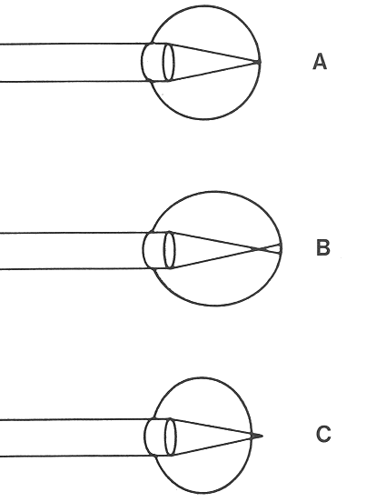
- Wrong focusing:
Due to various conditions, the optics of the eye and the shape of the eye may not
match. Consequently the image of the object formed by the optics of the eye may be
either in front or behind the retina, instead of being properly focused on the retina.
In either case, what the retina receives is a blurred image as it is out-of-focus.

Due to the aberration and diffraction, a perfect point light will have an imperfect
image of some finite size and some asymmetric shape. The 2D function representing
the shape of this imperfect image of a perfect point, called point-spread
function, is used to describe the distortion caused by the optics and its Fourier
transform is called the optical transform function, which can be used to predict
the image of any other non-point object, based on the assumption that the optical
system is a linear and shift-invariant system.
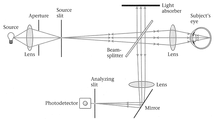
The aberrations of the optics of the eye can be studied by shining a light into
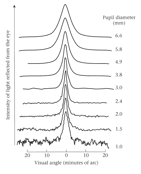
the eye and receiving the reflection from the retina. Campbell and Gubisch (1966)
used a signle line displayed on a monitor screen and recorded its reflection from
the observor's retina as a 1D funciton of space for different pupil diameters
ranging from 1.0 mm to 6.6 mm. In the ideal situation with no aberration or
distortion, the image reflected from the retina should also be a single line.
However, the actual retina reflection measured as a spatial function has the general
shape of a single peak (corresponding to the single line of incoming signal) whose
width increases as the pupil diameter increases, as larger pupil diameter will
introduce more aberrations to the opitcs. However, on the other hand, the larger
pupil diameter allows more light to pass and thereby higher signal-to-noise ratio.


The recorded function which represents the light going through the optics twice (in
and out) is further corrected to obtain the line-spread function, the actual image of
the single line formed on the retina. This line-spread function has similar shape
as the measured function with a single peak whose width also increases as the pupil
diameter increases, reflecting the fact that larger aperture of the optical system
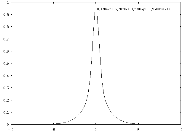
will cause poorer focusing. Westheimer (1986) propsed a formular to desribe this
line-spread function at pupil diameter of 3 mm:
h(x)=0.47e-3.3x2+0.53e-.93|x|

Figure:
Line-spread function of eye
|


Next: About this document ...
Up: The Eye
Previous: The optics of the
Ruye Wang
1999-09-22
![]() as the cross section (where J1 is the first order
Bessel function and r is the distance from the center).
as the cross section (where J1 is the first order
Bessel function and r is the distance from the center).


