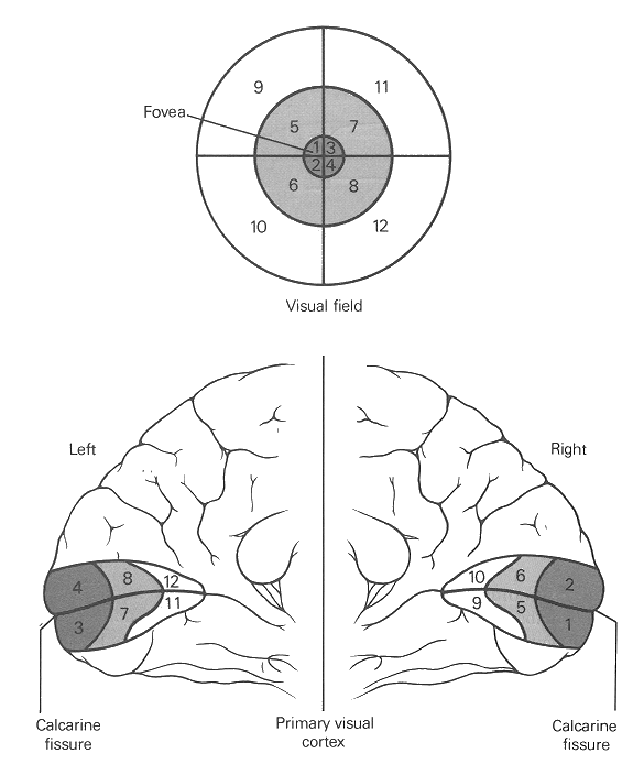
In lower visual areas (e.g., V1 through V5) the neurons are organized in an orderly fashion called topograpphic or retinotopic mapping, in the sense that they form a 2D representation of the visual image formed on the retina in such a way that neighboring regions of the image are represented by neighboring regions of the visual area. However, the retinotopic representation in the cortical areas is distorted. The foveal area is represented by a relatively larger area in V1 than the peripharal areas.
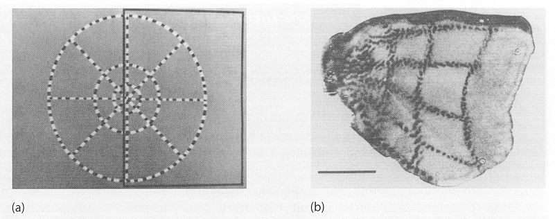
The retinotopic mapping is clearly demonstrated by an experiment (Tootell et al, 1982) as shown here:
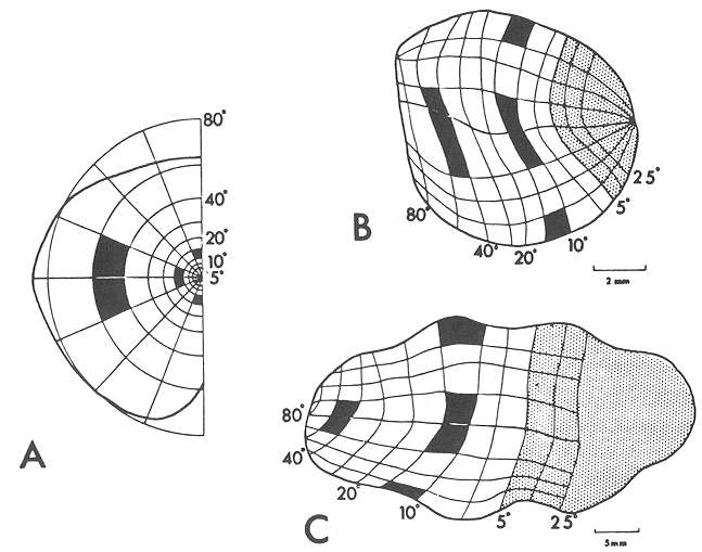
The following figure (Connolly and Van Essen 1984) shows the mapping of the visual field (A) on the LGN (B) and the striate cortex (C) in monkey. Note that the representation of the central 5 degrees (shaded areas) in the visual field occupies about 40 % of the cortex.
Also note that in this mapping, the upper region of the image is represented by the lower part of the V1 area, and the left side of the image is represented by the V1 area of the right hemisphere, and vice versa, as illustrated here: