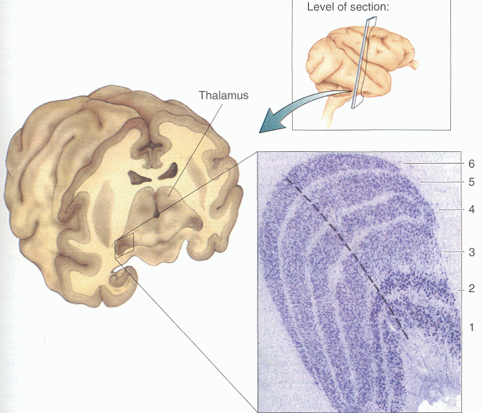
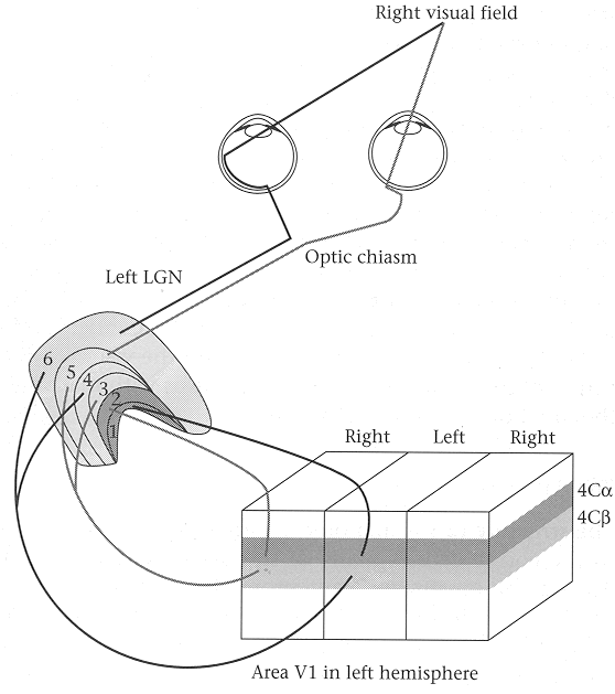
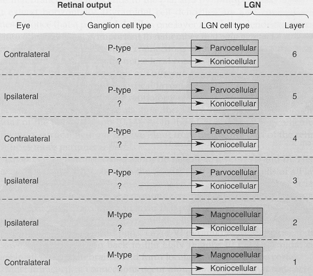
Lateral Geniculate Nucleus (LGN) is locateed in the most lateral, inferior region of the thalamus. It is composed of six layers, with the ipsilateral eye projecting to layers 2,3 and 5 and the contralateral eye projecting to layers 1,4 and 6. The two ventral layers, layers 1 and 2, contain the larger magnocellular cells receiving projections mainly from the magnocellular (parasol) ganglion cells, while the four dorsal layers, layers 3 through 6, contain smaller parvocellular cells receiving projections from mainly the parvocellular (midget) ganglion cells.


Each of the six layers has two sublayers. The upper (dorsal) one is the principal sublayer. The lower (ventral) one is the koniocellular sublayer (Greek konis means dust), as the cell bodies in the lower sublayers are smaller than those in the upper sublayers.
The projections from the retina ganglion cells to the LGN are retiontopic and registered so that a given point in the visual field is represented by the same point of all LGN layers along the same projection line perpendicular to the layers.
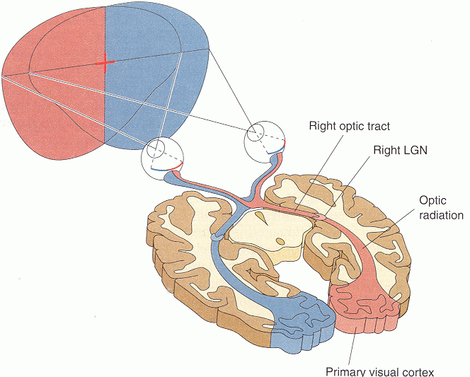
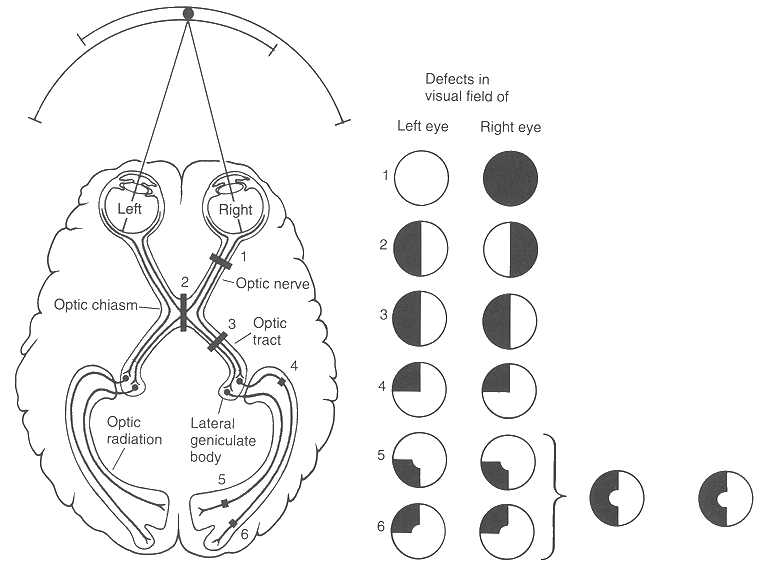
This figure shows visual deficits produced by lesions at various points in the visual pathway, shown as dark areas in the visual field maps. Deficits in the visual field of the left eye, for example, represent what one would see with the right eye closed, not deficits of the left visual hemifield of the brain.